Endobronchial Ultrasound (EBUS)
Endobronchial Ultrasound (EBUS) in Glendale, CA
Advanced Imaging for Accurate Lung and Lymph Node Diagnosis

Get Accurate Lung Diagnostics with Expert Endobronchial Ultrasound (EBUS) in Glendale, CA
If you’re facing persistent respiratory symptoms or have abnormal imaging results, our state-of-the-art pulmonary care center in Glendale, CA offers advanced Endobronchial Ultrasound (EBUS) to provide accurate, minimally invasive diagnostics. EBUS allows our specialists to visualize and biopsy lymph nodes and masses within the chest in real time—making it an essential tool for diagnosing conditions such as lung cancer, infections, and unexplained mediastinal abnormalities.
Our experienced team is proud to offer the trusted EBUS Glendale, CA patients depend on for precision, comfort, and results. With this cutting-edge procedure, we’re able to deliver earlier detection and more personalized treatment plans without the need for surgical intervention. Let us help you take the next step in your respiratory care with expert guidance and advanced diagnostic support.
Types of Endobronchial Ultrasound
Endobronchial ultrasound (EBUS) comes in two main types, each suited for specific diagnostic purposes:
1. Radial EBUS: This type is used to examine the outer walls of the lungs and surrounding tissues. It provides high-resolution images of lung masses, lymph nodes, and other structures that might be difficult to visualize with traditional methods.
2. Linear EBUS: A more advanced form, linear EBUS is equipped with a probe that can be used to guide a biopsy during the procedure, making it highly effective for staging cancer and diagnosing infections or other diseases. It is particularly helpful for collecting samples from difficult-to-reach areas of the lungs and nearby lymph nodes.
How Does an Endobronchial Ultrasound Work?
An EBUS involves the use of a bronchoscope, which is a flexible tube with a camera and an ultrasound probe at the tip. The bronchoscope is carefully inserted through the mouth and into the lungs. The ultrasound probe generates high-frequency sound waves that create detailed images of the lungs, airways, and surrounding lymph nodes. These images allow Dr. Ramyar Mahdavi to evaluate the structures and detect any abnormalities.
In addition to imaging, a linear EBUS can be used to guide a needle biopsy for further examination of tissue or lymph nodes, which helps with accurate diagnosis and treatment planning.
Why Would You Need an EBUS?
Endobronchial ultrasound is commonly recommended for patients who experience symptoms like persistent cough, chest pain, unexplained weight loss, or difficulty breathing. It is a key diagnostic tool for:
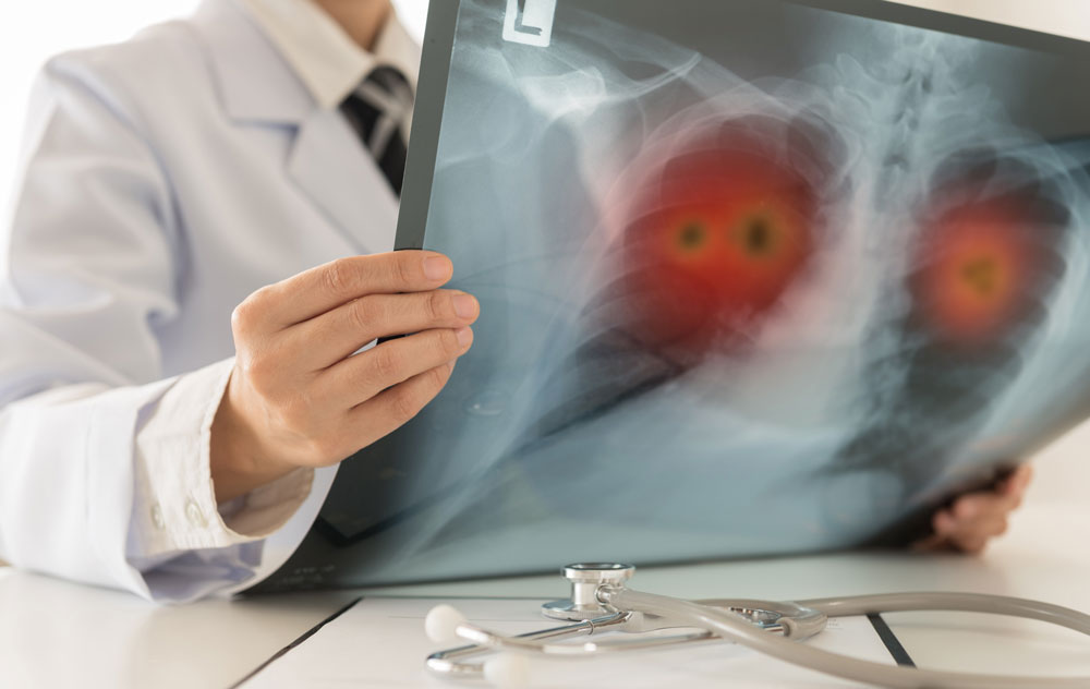
- Detecting Lung Cancer: Identifying the presence of tumors and staging lung cancer for better treatment planning.
- Investigating Lymph Node Involvement: Checking for enlarged lymph nodes that could indicate infection, inflammation, or cancer.
- Diagnosing Infections or Inflammation: Identifying causes of chronic cough or unexplained symptoms that might indicate tuberculosis, pneumonia, or other lung conditions.
- Biopsy Guidance: Assisting in obtaining tissue samples for biopsy when other diagnostic procedures may not be possible.
What Are the Benefits of an Endobronchial Ultrasound?
Endobronchial Ultrasound offers numerous advantages, including:
- Minimally Invasive: No need for surgery or large incisions, making recovery faster and easier.
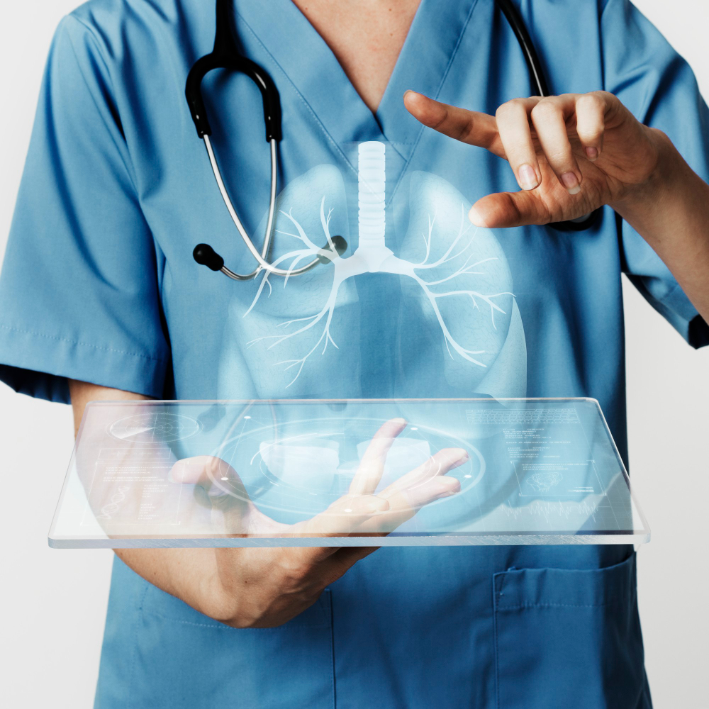
- Accurate and Detailed Imaging: Provides real-time, high-resolution images of the lungs and surrounding structures.
- Guided Biopsy: Enables precise biopsies of abnormal tissue, improving diagnostic accuracy.
- Early Detection: Allows for the detection of diseases like cancer and infections in their early stages, increasing the effectiveness of treatment.
- Outpatient Procedure: EBUS is typically performed on an outpatient basis, meaning you can return home the same day.
What Should I Expect During an Endobronchial Ultrasound?
An EBUS procedure is minimally invasive, but it’s important to understand the process:
- Preparation: You may be asked to fast for several hours before the procedure to ensure your stomach is empty.
- Sedation: To ensure your comfort, you will be given a mild sedative and local anesthesia to numb the throat. This procedure is performed under general anesthesia.
- Procedure: Dr. Mahdavi will insert the bronchoscope through the mouth and guide it into your airways. The ultrasound probe will capture detailed images while the bronchoscope provides real-time visuals of your lungs.
- Biopsy (if needed): If necessary, a needle biopsy may be performed to collect tissue samples for further testing.
- Recovery: The procedure typically takes about 30-60 minutes. Afterward, you’ll be monitored for a short time (usually an hour or two) before being allowed to go home.
What Are the Risks of an EBUS?
Endobronchial Ultrasound (EBUS) is generally safe, but like any medical procedure, there are some risks, including:
- Bleeding: Rare, but bleeding may occur if a biopsy is taken.
- Infection: A small risk of infection at the biopsy site.
- Discomfort: Temporary sore throat or coughing can occur post-procedure.
Why Choose Us for Your Endobronchial Ultrasound (EBUS) in Glendale, CA?
At Dr. Ramyar Mahdavi’s Glendale clinic, we provide expert, compassionate, and patient-focused care. Here’s why we’re the top choice for Endobronchial Ultrasound (EBUS):
- Expertise and Experience: Dr. Mahdavi is a highly trained interventional pulmonologist with years of experience in performing advanced diagnostic procedures like EBUS. His deep understanding of respiratory conditions ensures that you receive the most precise and effective care.
- State-of-the-Art Technology: We use the latest EBUS equipment, which allows us to obtain high-quality imaging and accurate biopsy results. This cutting-edge technology enhances our ability to detect and diagnose lung and airway issues early.
- Patient-Centered Care: We prioritize your comfort and well-being throughout the entire process. From your initial consultation to post-procedure follow-up, we make sure you feel informed, supported, and at ease.
- Minimally Invasive Approach: Our EBUS procedures are designed to be minimally invasive, reducing recovery time and helping you return to your normal routine more quickly compared to traditional surgery.
- Comprehensive Diagnosis and Treatment Plans: After your EBUS procedure, Dr. Mahdavi works with you to discuss the results and develop a personalized treatment plan, whether you need further tests or immediate care for your condition.
- Convenient Location in Glendale, CA: Our clinic is centrally located, making it easy for you to access our services. We provide convenient appointment scheduling and strive to minimize wait times.
Contact Dr. Ramyar Mahdavi for Your Endobronchial Ultrasound (EBUS) Consultation in Glendale, CA
If you’re experiencing respiratory symptoms or need an evaluation for lung or airway issues, Dr. Ramyar Mahdavi is here to help. Schedule your consultation today and take the first step toward better lung health.
Frequently Asked Questions (FAQ’s)
No, Endobronchial Ultrasound (EBUS) is not painful. The procedure is performed under general anesthesia, so you will be comfortable throughout. Most patients experience minimal discomfort, such as a mild sore throat or hoarseness afterward, but the procedure itself is generally painless.
The EBUS procedure itself typically lasts 30 to 60 minutes. Once completed, the tissue samples taken during the procedure will be sent for analysis. Depending on the complexity of the tests, the results are usually available within a few days to a week. Dr. Mahdavi will discuss the findings with you and explain what they mean for your treatment options.
Yes, preparation is important. You will be asked to fast for at least 6-8 hours before the procedure to ensure your stomach is empty. This reduces the risk of complications such as aspiration (inhaling food or liquid into the lungs). Dr. Mahdavi’s office will provide detailed instructions on how to prepare and whether any of your medications need to be adjusted.
While EBUS is generally safe, there are some risks involved, as with any medical procedure. These include mild bleeding, infection, or a sore throat. However, these risks are uncommon, and Dr. Mahdavi takes all necessary precautions to ensure the procedure is as safe as possible.
Yes, since EBUS is performed under general anesthesia, you will need someone to drive you home afterward. The sedation may leave you feeling groggy or drowsy, so it’s important to have a friend or family member with you to ensure your safety.
EBUS is highly accurate and a valuable diagnostic tool for evaluating lung conditions. It provides real-time imaging of the lungs and surrounding structures, allowing Dr. Mahdavi to guide tissue sampling with precision. It is particularly effective for diagnosing lung cancer, infections, and conditions involving the lymph nodes in the chest.